Yannick Schwab
Team Leader and Head of Electron Microscopy Core Facility
Schwab Team
ORCID: 0000-0001-8027-1836
EditThe facility provides advanced expertise in electron microscopy, from sample preparation to image analysis, for a wide variety of biological samples.
Team Leader and Head of Electron Microscopy Core Facility
Schwab Team
ORCID: 0000-0001-8027-1836
EditThe EMCF provides advanced expertise in cellular electron microscopy: from sample preparation to image analysis and for a large variety of biological samples ranging from bacterial cells to small multicellular organisms. Our main focus is on ultrastructural analysis in 2D and 3D and in correlative light and electron microscopy (CLEM). Our staff can help you define optimal experimental conditions for your project.
We are involved in method development, which is heavily connected to our collaborations with groups here at the EMBL. An example of a this is the on-section, correlative light and electron microscopy (CLEM) technique, which tracks the signal of fluorescent proteins in resin sections with high precision. We have published a detailed step-by-step video tutorial on this method and we are hosting an international course covering the methodology every second year.
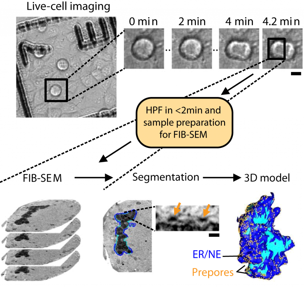
Together with the Ephrussi, Jechlinger, Leptin and Mahamid groups we have developed high-precision targeting methods for volume electron microscopy (Ronchi et al., 2021). The strategy relies on fluorescence preservation during sample preparation and targeted trimming guided by confocal maps of the fluorescence signal in the resin block. Laser branding is used to create landmarks on the block surface to position the FIB-SEM acquisition. Click here to see the video of the full workflow.
In 2019 we have also developed pipeline for automated TEM (Schorb et al., 2019). With this technique, one is able to image many individual cells within a tissue or culture in a short period of time, which can later be used for statistical analysis. Here we drastically reduce the beam time by imaging the individual cells and not the whole section.

Please note that single-particle and tomography cryo-EM for structural biology projects are offered through the cryo-EM service platform and the EMBL IC.
Profiling cellular diversity in sponges informs animal cell type and nervous system evolution.
Musser JM, Schippers KJ, Nickel M, Mizzon G, Kohn AB, Pape C, Ronchi P, Papadopoulos N, Tarashansky AJ, Hammel JU, Wolf F, Liang C, Hernández-Plaza A, Cantalapiedra CP, Achim K, Schieber NL, Pan L, Ruperti F, Francis WR, Vargas S, Kling S, Renkert M, Polikarpov M, Bourenkov G, Feuda R, Gaspar I, Burkhardt P, Wang B, Bork P, Beck M, Schneider TR, Kreshuk A, Wörheide G, Huerta-Cepas J, Schwab Y, Moroz LL, Arendt D. Science. 2021 Nov 5;374(6568):717-723.
High-precision targeting workflow for volume electron microscopy.
Ronchi P, Mizzon G, Machado P, D’Imprima E, Best BT, Cassella L, Schnorrenberg S, Montero MG, Jechlinger M, Ephrussi A, Leptin M, Mahamid J, Schwab Y. J Cell Biol. 2021 Sep 6;220(9):e202104069.
Integrative Imaging Reveals SARS-CoV-2-Induced Reshaping of Subcellular Morphologies.
Cortese M, Lee JY, Cerikan B, Neufeldt CJ, Oorschot VMJ, Köhrer S, Hennies J, Schieber NL, Ronchi P, Mizzon G, Romero-Brey I, Santarella-Mellwig R, Schorb M, Boermel M, Mocaer K, Beckwith MS, Templin RM, Gross V, Pape C, Tischer C, Frankish J, Horvat NK, Laketa V, Stanifer M, Boulant S, Ruggieri A, Chatel-Chaix L, Schwab Y, Bartenschlager R. Cell Host Microbe. 2020 Dec 9;28(6):853-866.
Software tools for automated transmission electron microscopy.
Schorb M, Haberbosch I, Hagen WJH, Schwab Y, Mastronarde DN.
Nat Methods. 2019 Jun;16(6):471-477.


If you would like to apply as a visitor in the EMCF, please contact us at emcf@embl.de including a short overview of your project. We’ll discuss the feasibility of your project internally and in case of a positive evaluation we will contact you for a more detailed meeting over the phone, via Zoom or video conference.
The EMCF currently participates in several initiatives supporting visitor access:
The EMCF is part of the EMBL node in Euro-BioImaging, together with the Advanced Light Microscopy Facility (ALMF) at EMBL Heidelberg and the Mesoscopic Imaging Facility (MIF) at EMBL Barcelona. The EMBL node offers open access to a broad range of state-of-the-art technologies in biological imaging for life scientists.
The Fellowships support travel and accommodation costs of young scientists visiting EMBL Core Facilities at the different EMBL sites.
EMBL Corporate Partnership Programme Fellowships
The Corporate Partnership Programme continues to provide financial support for scientists to train and collaborate in EMBL research groups and core facilities.
EMBO Core Facility Fellowships
EMBO is currently offering fellowships to support international exchange and training of staff working in a core facility.

